Chapter Five

Lichen Identification Guide

From field observations and hand-lens work to chemical spot tests and lab chromatography, a systematic approach to putting a name on the lichen in front of you.

Updated October 2026: added an “is it a lichen?” check, a field key from growth form to genus, a spot-test quick reference, side-by-side look-alike tables and a photo checklist for getting an ID online.

Why Identification Matters

With roughly 20,000 described species worldwide, lichen identification is both deeply rewarding and genuinely challenging. Many species look nearly identical to the naked eye but differ in their chemistry. Others change appearance dramatically between wet and dry conditions. A systematic approach (combining visual observation, chemical tests, and sometimes lab work) is essential for reliable identification.

The process below moves from the simplest observations anyone can make in the field to advanced techniques used by professional lichenologists. You can often narrow a specimen to genus with just the first few steps, but species-level identification frequently requires chemistry.

On This Page

Is It a Lichen at All?

Several things on bark and rock are regularly mistaken for lichens. Rule them out first:

What you seeProbablyHow to tell
Tiny green leaves on stems, soft cushions or carpets Moss (a plant) Under a lens you can see individual leaves on a stem. Lichens have no leaves or stems; they are crusts, lobes, cups or branches.
Thin, bright green film or stain on bark, fences or walls, often on the damp side Free-living green algae A uniform green coating with no lobes, cups, discs or powdery granules. A lichen crust usually has a definite edge, texture or fruiting bodies.
Orange to rusty film on bark, rock or mortar Trentepohlia, a free-living orange alga A powdery film with no lobes or discs. Orange lichens such as Xanthoria have lobes or fruiting discs and turn K+ purple.
Soft, pale grey to blue-grey powder on shaded bark or rock overhangs A dust lichen (Lepraria) A lichen made entirely of soredia-like granules, with no cortex. Usually paler and greyer than an algal film; L. incana glows blue-white under UV.
Brackets, gills, jelly blobs on dead wood that appear after rain and rot away A non-lichen fungus Scratch a lichen and you usually see a thin green layer of algae just under the surface; other fungi have no such layer.

The 8-Step Visual ID Process

Before reaching for reagents or a microscope, careful observation with a hand lens (10x) will take you surprisingly far. Work through these eight steps in order for every specimen.

Step Observation Key Questions Narrows To
1 Growth Form Crustose, foliose, fruticose, squamulose, gelatinous, or leprose? Major group
2 Color Green, gray, yellow, orange, brown, black? Note both wet and dry color. Order / Family
3 Substrate On bark (corticolous), rock (saxicolous), soil (terricolous), or wood (lignicolous)? Ecological group
4 Lobe Shape Broad or narrow lobes? Rounded or pointed tips? Flat or ascending margins? Genus
5 Reproduction Soredia, isidia, apothecia, perithecia, or lobules present? Species group
6 Underside Black, white, brown, or tan? Rhizines simple or branched? Veins visible? Genus / Species
7 Special Features Cilia, pseudocyphellae, cyphellae, tomentum, pruina, or lirellae? Species
8 Geography & Habitat Region, elevation, forest type, microhabitat, exposure? Confirms ID

Step-by-Step in Detail

Growth Form

This is always your starting point. Is the lichen a flat crust fused to the surface (crustose), a leafy structure you can peel up at the edges (foliose), a three-dimensional shrubby or hanging form (fruticose), overlapping scales (squamulose), a jelly-like mass (gelatinous), or a powdery dusting with no visible structure (leprose)? Growth form alone eliminates the vast majority of possible species.
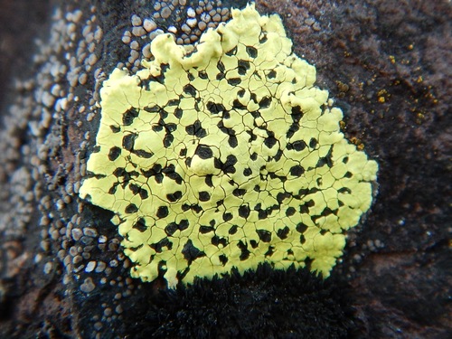
Crustose lichen on rock Crustose
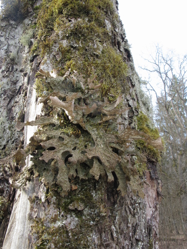
Foliose lichen with leafy lobes Foliose
Fruticose lichen hanging from branch Fruticose
Squamulose lichen with overlapping scales Squamulose
Gelatinous lichen Gelatinous
Crustose lichen with lirellae Leprose / Crustose

Color

Record the color carefully, and note whether the specimen is wet or dry, since many lichens change color dramatically with moisture. Dry gray-green lichens often become vivid green when wet. Bright yellows and oranges usually indicate specific chemical compounds (usnic acid, anthraquinones) that narrow identification immediately.

Substrate

What the lichen is growing on matters enormously. Many species are strict specialists: Xanthoria parietina favors nutrient-enriched bark and rock, while Rhizocarpon geographicum grows exclusively on siliceous rock. Note the specific tree species if on bark, and rock type (limestone vs. granite) if saxicolous.

Saxicolous lichen on rock Saxicolous (rock)
Corticolous lichen on bark Corticolous (bark)
Terricolous lichen on soil Terricolous (soil)
Lignicolous lichen on wood Lignicolous (wood)

Lobe Shape

For foliose lichens, lobe morphology is critical. Are lobes broad (over 5 mm) or narrow? Are the tips rounded, truncate, or pointed? Do the margins curl up, lie flat, or curl under? Are the lobes tightly appressed to the substrate or loosely ascending? These details separate genera that look superficially similar.

Reproductive Structures

Look for apothecia (open disc-shaped fruiting bodies), perithecia (flask-shaped bodies embedded in the thallus), soredia (powdery granules), isidia (finger-like outgrowths), or lobules (tiny lobe-like propagules). The presence or absence of these structures is one of the most important species-level characters.
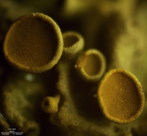
Close-up of orange cup-shaped apothecia on Xanthoria parietina Apothecia
Perithecia on Verrucaria Perithecia
Soredia in cracks of Parmelia sulcata Soredia
Isidia on Parmelia saxatilis Isidia
Podetia of Cladonia cristatella Podetia

Underside Examination

Gently lift a lobe edge to examine the lower surface. Is it black, brown, white, or tan? Are there rhizines (root-like attachment structures), and if so, are they simple, squarrose (brush-like), or dichotomously branched? Some genera have distinctive undersurface features: Peltigera has raised veins, and Sticta has round pores called cyphellae.
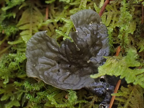
Peltigera showing veined underside and rhizines Rhizines & veins (Peltigera)
Sticta showing cyphellae Cyphellae (Sticta)
Punctelia showing pseudocyphellae Pseudocyphellae (Punctelia)

Special Features

Look for cilia (hair-like projections from lobe margins), pseudocyphellae (white dots or lines on the upper surface that are pores in the cortex), tomentum (a fuzzy felt-like covering), or pruina (a frosty white coating). These micro-characters often clinch the identification when everything else is ambiguous.

Geography & Habitat

Where you are matters. Many lichens have restricted geographic ranges, and distribution is a valid identification character. Note your region, elevation, forest type (old-growth vs. secondary), aspect (north-facing vs. south-facing), and proximity to pollution sources. A lichen that looks like Lobaria pulmonaria but grows near a highway probably is not — it requires clean air.
Soredia vs. Isidia — The Critical Distinction

Both are asexual reproductive structures, but they differ fundamentally and separating them is one of the most important skills in lichen identification.

Soredia on Parmelia sulcata

Soredia

Powdery granules erupting from breaks in the cortex (called soralia). Loose bundles of algal cells wrapped in fungal hyphae, with no cortex of their own. Look dusty or granular under a hand lens.

Isidia on Parmelia saxatilis

Isidia

Finger-like, coral-like, or cylindrical outgrowths from the thallus surface. Crucially, they have their own cortex: a smooth, intact outer layer. Look like tiny organized projections, not like dust.

Many species pairs are separated solely by this character. For example, Parmelia sulcata has soredia in its cracks, while Parmelia saxatilis has isidia. They are otherwise nearly identical.

Field Key: From Growth Form to Genus

Start with the growth form, then follow the colour and substrate to a short list. Each row gives the features you can actually see (or test) and the common lichens that match them in North America and Europe. A photo-based match is usually safe to genus; confirming the species often needs the spot test listed, or a regional guide.

1. Hanging or shrubby (fruticose)

Colour & shapeWhereCheckLikely
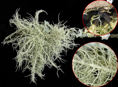
Pale yellow-green, round branchesTree branchesPull a strand: white elastic central cordUsnea (old man’s beard); one very long strand with short, even side branches = Dolichousnea longissima
Pale yellow-green, round, stiff hair-like strandsConifer branchesNo central cordAlectoria
Brown to black hair-like tanglesConifer branchesNo central cordBryoria (horsehair lichen)
Flat straps, same colour on both sides, firmBark, twigsNo cord; often marginal soraliaRamalina; lace-like nets in coastal California = R. menziesii
Soft flat straps, green-grey above, white belowBark, twigsAll spot tests negativeEvernia prunastri (oakmoss)
Grey straps, underside darkening to black, scurfy with isidiaConifer barkCortex K+ yellowPseudevernia furfuracea
Brilliant chartreuse-yellow tuftsConifers and old wood, western North AmericaColour alone is distinctiveLetharia vulpina (wolf lichen)
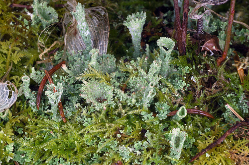
Grey-green or yellowish cups, sticks or red-tipped stalksSoil, logs, stumpsHollow stalks rising from small scalesCladonia: see the cups and sticks table
Grey or yellowish, much-branched matsGround in boreal forest, heath, tundraK and P testReindeer lichens: see the reindeer table
Olive to chestnut-brown channelled lobes with spiny edgesHeath, tundra, mountain soilWhite pseudocyphellae on the undersideCetraria islandica (Iceland moss)

2. Leafy, with a separate underside (foliose)

Colour & shapeWhereCheckLikely
Grey with a net of white linesBark, rockSoredia or isidia in the net? Underside blackParmelia sulcata (soredia) or P. saxatilis (isidia)
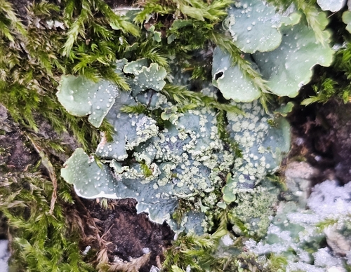
Grey with scattered white dotsBarkMedulla C+ red; underside tan to brownPunctelia
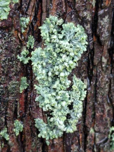
Yellow-green, broad wrinkled lobes, no white dotsBark, especially oakMedulla P+ orange-red, C−Flavoparmelia caperata (common greenshield)
Yellow-green, tightly attachedRockAlways on rockXanthoparmelia (rock shields)
Grey, broad lobes with black hairs (cilia) on the marginsBarkUnderside black with a bare brown edgeParmotrema
Grey, puffy hollow lobes, black underside, no rhizinesTwigs, conifer barkSqueeze a lobe: it is hollowHypogymnia physodes
Small pale grey to white rosettes, narrow lobesTwigs, enriched bark, wallsCortex K+ yellowPhyscia; see the rosette table
Orange to yellow-orangeBark, rock, concreteK+ purpleXanthoria, Xanthomendoza, Rusavskia
Tiny lemon-yellow lobes under 1 cmRoadside tree barkK−Candelaria concolor
Large lobes, green when wet, with dark “freckles”Moss and soilCephalodia on the upper surfacePeltigera aphthosa
Large grey-brown lobes, veined felty undersideMoss, soil, logsRaised veins and long rhizines belowPeltigera (dog lichens)
Large green-brown lobes with a network of ridgesOld trees in humid forestLooks like lung tissueLobaria pulmonaria
Leathery, held by one central pointRockSingle central holdfast (umbilicus)Umbilicaria (rock tripe)

3. Crust fused to the surface (crustose and leprose)

Colour & shapeWhereCheckLikely
Yellow-green patches outlined in blackSiliceous rock, uplandsBlack prothallus between areolesRhizocarpon geographicum (map lichen)
Egg-yolk yellow granulesRock, concrete, postsK−Candelariella vitellina
Orange crust or lobed rosetteRock, wallsK+ purpleCaloplaca and relatives, Rusavskia elegans
Yellow-grey rosettes with lobed edges and crowded discsWalls, pavements, gravestonesDiscs paler than the thallusProtoparmeliopsis muralis
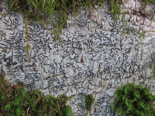
Grey-white crust with black, script-like linesSmooth barkLirellaeGraphis scripta
Thin white crust like whitewashShaded barkK+ yellow turning redPhlyctis argena
Matt black bandSeashore rock above high tideCracked, with tiny black dotsHydropunctaria maura (black tar lichen)
Pink balls on short white stalksBare acid soil, road cutsStalked, round headsDibaeis baeomyces
Mint-green crust with flat pink discsRotting logs, peatDiscs unstalkedIcmadophila ericetorum
Grey-green crust ringed with redBark, southeastern USRed cottony rim, white bandHerpothallon rubrocinctum (Christmas lichen)
Pale grey to blue-grey powder onlyShaded bark, overhangsNo cortex, no discsLepraria (dust lichens)

4. Rubbery or jelly-like when wet (gelatinous)

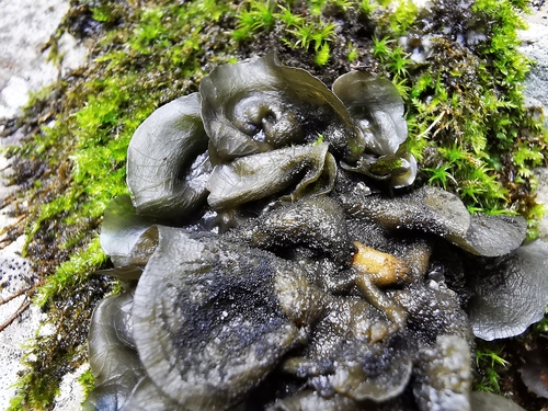
Dark olive to black, brittle when dry, swelling and translucent within minutes of rain. These are cyanobacterial lichens: Collema has no cortex and becomes thick and pulpy when wet; Leptogium and Scytinium have a cortex one cell thick and are usually thinner and wrinkled. Several small species are now split between these genera, and a microscope section is the reliable test.

Chemical Spot Tests

Many lichen species are visually identical but differ in their secondary chemistry. Five reagent tests (each targeting different compound classes) let you distinguish them in the field. Apply reagents to the cortex or scratch through to the medulla, depending on which layer you need to test.

K

K Test — Potassium Hydroxide

Apply a small drop of 10% KOH solution to the cortex or exposed medulla. Watch for a color change: K+ yellow indicates atranorin; K+ yellow turning red indicates salazinic acid; K+ orange-brown suggests norstictic acid. K− (no reaction) is equally informative. This is the single most useful spot test and the one to learn first.

C

C Test — Calcium Hypochlorite (Bleach)

Apply diluted household bleach to the cortex or medulla. C+ pink or red indicates gyrophoric acid or lecanoric acid. Many lichens are C− (no reaction), which is also a useful data point. The C test is especially important for crustose species where K alone is insufficient.

KC

KC Test — Sequential Application

Apply K first, then immediately apply C to the same spot. The combined reaction may differ from either reagent alone. KC+ pink or red-orange detects alectoronic acid and physodic acid, compounds that are invisible to K or C individually. Always record K and C results separately before testing KC.

P

P Test — Paraphenylenediamine

Apply P reagent to the medulla (scratch through the cortex first). P+ orange or red indicates fumarprotocetraric acid or protocetraric acid. These are among the most common lichen compounds and P is essential for distinguishing many look-alike species.

Safety warning: Paraphenylenediamine is toxic and a known carcinogen. Always wear gloves and avoid skin contact. Work in a well-ventilated area. Many field lichenologists use P dissolved in ethanol and apply it with a fine brush or cotton swab. Dispose of waste properly.

UV

UV Fluorescence — Long-wave 365 nm

Some lichen compounds fluoresce under long-wave ultraviolet light. Blue-white, orange, or yellow fluorescence can indicate divaricatic acid, squamatic acid, and other compounds. UV is non-destructive, requires no reagents, and is especially useful in the field after dark. A portable 365 nm UV flashlight is an inexpensive but powerful addition to any lichen toolkit.

Spot-Test Quick Reference

The reactions you are most likely to see on common species, and what they point to. Results apply to the layer named: scratch through the cortex with a blade to test the white medulla.

ReactionLayerUsual causeCommon examples
K+ yellowCortexAtranorinPhyscia, Parmelia, Punctelia, Pseudevernia, Cladonia rangiferina
K+ purple to crimsonCortex, discsAnthraquinones (parietin)Xanthoria, Xanthomendoza, Rusavskia, Caloplaca, Teloschistes
K+ yellow, slowly turning redMedulla or thallusSalazinic or norstictic acidParmelia sulcata and P. saxatilis (salazinic); Phlyctis argena (norstictic)
C+ red (fleeting)MedullaLecanoric acid (gyrophoric acid gives C+ pink to rose)Punctelia rudecta, Flavopunctelia
KC+ yellowCortexUsnic acidAlectoria, Cladonia stellaris
P+ red or orange-redMedulla, podetiaFumarprotocetraric or protocetraric acidCladonia pyxidata, C. chlorophaea, C. fimbriata, C. rangiferina, Cetraria islandica, Flavoparmelia caperata
UV+ white or blue-whiteThallusSquamatic, perlatolic and other acidsLepraria incana, one chemotype of Cladonia squamosa, Icmadophila, some C. stellaris
All negative–No reactive compoundsCandelaria concolor, Evernia prunastri, Hydropunctaria maura

Getting reliable results.

Use a tiny drop on a small spot and watch it through a hand lens as it goes on. C and KC reactions can flash and fade within about a second, so look at the moment of application. Some K reactions develop: salazinic acid goes yellow first and turns red over the next few seconds. Household bleach works as C reagent but weakens quickly, so replace it often and keep it in a dark bottle. K (10% potassium hydroxide) is caustic, and P stains skin and is toxic. Always note negative results too, since “K−” is as useful as “K+”.

Thin-Layer Chromatography (TLC)

When spot tests are ambiguous or you need to identify the exact compound present, thin-layer chromatography is the gold standard. TLC separates individual chemical compounds from a lichen sample and compares them to known standards, giving definitive chemical identification.

How TLC Works

A small fragment of lichen is dissolved in acetone or another organic solvent and spotted onto a glass plate coated with silica gel. The plate is placed upright in a developing chamber containing a carefully calibrated solvent system. As the solvent wicks upward by capillary action, different compounds travel at different rates depending on their polarity and molecular weight. The result is a series of separated spots, each representing a single compound.

By comparing the position (Rf value) and color of each spot against known standards run on the same plate, the lichenologist can identify the exact compounds present. This is how the more than 800 known lichen secondary compounds have been catalogued, and it remains the definitive method for chemical identification when spot tests leave doubt.

When to use TLC: TLC is most valuable when two species are morphologically identical and differ only in chemistry, so-called chemical species or chemotypes. It is also essential when publishing new species descriptions, as chemical data is now considered a required component of lichen taxonomy.

Key Look-Alike Pairs

These commonly confused species pairs illustrate why careful observation and chemistry matter. Learning to separate them will sharpen your identification skills across the board.

Parmelia sulcata showing soredia
vs
Parmelia saxatilis showing isidia

Parmelia sulcata

Hammered Shield Lichen

Gray-green foliose lichen with a distinctive network of ridges (reticulate pattern) on the upper surface. Soredia develop in the cracks of this network. Look for powdery, granular material filling the grooves. Very common on bark and fences throughout the Northern Hemisphere.

K+ yellow • Soredia in cracks

Parmelia saxatilis

Crudely Wrinkled Shield Lichen

Nearly identical gray-green foliose lichen with the same reticulate upper surface. But instead of soredia, it bears cylindrical isidia: small, cortex-covered outgrowths. Under a hand lens, the surface looks bumpy and organized rather than dusty.

K+ yellow • Isidia (not soredia)

Usnea hanging from branch
vs
Bryoria hanging from branch

Usnea

Old Man's Beard

Pale green to gray-green hanging fruticose lichen. The diagnostic test: gently stretch a branch. An elastic white central cord will be revealed inside. This is unique to Usnea and present in every species of the genus. Color is typically pale yellowish-green. Contains usnic acid.

Elastic central cord present • Pale green

Bryoria

Horsehair Lichen

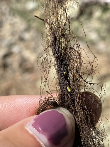
Dark brown to black hanging fruticose lichen that superficially resembles dark Usnea. The key difference: no central cord. When you pull a branch apart, it simply snaps or separates into fibers. There is no elastic white strand inside. Generally darker in color than Usnea.

No central cord • Dark brown to black

Cladonia chlorophaea with mealy soredia
vs
Cladonia pyxidata with corticate squamules

Cladonia chlorophaea

Mealy Pixie-Cup

Forms small trumpet-shaped cups (podetia) rising from a base of basal squamules. The inside and outside of the cups are covered in fine, granular soredia giving them a mealy, farinose texture. The cups appear powdery and slightly rough under magnification.

Cups sorediate (mealy) • P+ red

Cladonia pyxidata

Pebbled Pixie-Cup

Very similar trumpet-shaped cups, but instead of soredia, the surfaces are covered in corticate squamules: tiny, flattened, scale-like structures with a proper cortex. Under a lens, the cups look rough and scaly rather than powdery. A classic soredia-vs-cortex distinction.

Cups squamulose (scaly) • Corticate

Physcia adscendens with hooded soralia
vs
Physcia close-up showing lobe details

Physcia adscendens

Hooded Rosette Lichen

Small gray-white foliose lichen with ascending lobe tips. The soralia are hood-shaped (helmet-shaped): the lobe tip curls over to form a little hood with soredia produced on the inner, protected surface. Cilia present along lobe margins.

Hood-shaped soralia • Tips curl over

Physcia tenella

Resembles P. adscendens

Very similar gray-white foliose lichen, but the soralia are lip-shaped rather than hooded. The lobe tips bend back slightly to expose soredia on an open, lip-like surface rather than enclosing them in a hood. Cilia also present. Requires a hand lens to separate reliably from P. adscendens.

Lip-shaped soralia • Tips bend back

Evernia prunastri with white underside
vs
Ramalina farinacea, same color both sides

Evernia prunastri

Oakmoss

Soft, strap-shaped fruticose lichen with distinct upper and lower surfaces. The upper surface is gray-green while the underside is conspicuously white. This two-toned appearance is the quickest way to recognize Evernia. Used extensively in the perfume industry.

White underside • Two-toned

Ramalina farinacea

Farinose Cartilage Lichen

Also strap-shaped and fruticose, but the same color on both sides: a pale yellowish-green throughout. No distinct upper/lower differentiation. Often bears marginal or laminal soredia along the branches. Firmer and more cartilaginous than the soft straps of Evernia.

Same color both sides • Uniform

Look-Alike Comparison Tables

The groups that cause the most confusion, side by side. Read across a row for the features that separate each lichen from its neighbours. Each feature is something you can see with a 10× lens or check with a single spot test.

Beards and straps: Usnea vs Ramalina vs Evernia and relatives

LichenBranchesColourInside / undersideOther clues
UsneaRound in sectionPale yellow-green to grey-green; some species reddishWhite elastic central cordOften with soredia, isidia or tiny side spines; cortex usnic acid
Dolichousnea longissimaOne very long axis with short, even side branchesPale yellow-greenCentral cordStrands to several metres; humid old forest
RamalinaFlat or angular straps (a few species round)Same pale yellowish-green on both sidesNo cord; firm, cartilaginousSoralia on margins or surface in sorediate species
Evernia prunastriSoft, flat, forking strapsGreen-grey aboveWhite underside, no cordSoralia start as discrete spots on ridges and margins; all spot tests negative
Evernia mesomorphaAngular, flabby, ridgedPale yellow-green all roundNo white undersideCoarse soredia on the ridges
Pseudevernia furfuraceaFlat straps forking in one planeGrey aboveUnderside white to pinkish when young, blackening with ageIsidia; no rhizines; cortex K+ yellow
Alectoria sarmentosaRound, hair-like, stiffPale yellowish-green; tips may blackenNo cordWhite pseudocyphellae on the branches; cortex KC+ yellow
BryoriaRound, hair-likeBrown to blackNo cordNever KC+ yellow

Grey and green shields: Parmelia vs Punctelia vs Flavoparmelia

LichenColourWhite markingsPropagulesUndersideSpot test
Parmelia sulcataGrey to blue-greyNet of white lines (reticulate pseudocyphellae)Soredia in the netBlack, rhizines simple to bottle-brushMedulla K+ yellow→red
Parmelia saxatilisGrey, browner tipsNet of white linesIsidia, no sorediaBlack, rhizines simpleMedulla K+ yellow-orange→red
Punctelia rudectaGrey to blue-greyScattered white dotsIsidiaPale tan to brownMedulla C+ red
Punctelia subrudecta groupGreyWhite dotsSorediaPale brownMedulla C+ red
Flavoparmelia caperataPale yellow-greenNoneCoarse granular soredia in pustulesBlack, brown at the edgeMedulla P+ orange-red, C−
FlavopuncteliaYellow-greenWhite dots (minute in F. soredica)Soredia–Medulla C+ red
ParmotremaGreyUsually noneSoredia on lobe edgesBlack with a bare brown marginLobes broad, often with black cilia

Small rosettes on twigs and walls: Physcia vs Xanthoria and other K-test groups

These often grow together on nutrient-rich bark. One drop of K sorts most of them: grey ones that turn yellow are Physcia; orange ones that turn purple are Xanthoria and relatives; yellow ones that do nothing are Candelaria or Candelariella.

LichenColourLobes & featuresUndersideK test
Physcia (e.g. P. adscendens, P. tenella)Pale grey to whiteNarrow lobes; hooded (adscendens) or lip-shaped (tenella) soralia; marginal ciliaPaleCortex K+ yellow
Physcia stellarisPale greyStar-shaped rosettes, no soredia, many black-disced apotheciaPaleCortex K+ yellow
PhaeophysciaGrey-brown to dark greySmall lobes, never frostedUsually black; rhizines black with white tipsK−
PhysconiaGrey-brownLobe tips often frosted white (pruinose)Black bottle-brush rhizines forming a fringeK−
Xanthoria parietinaOrange to yellow-orange; grey-green in deep shadeBroad, flat lobes; abundant orange discsPale, with few rhizinesK+ purple
Xanthomendoza fallaxOrange-yellowSmall rosettes with hood-shaped soralia; discs rarePaleK+ purple
Rusavskia elegansIntense orange-redNarrow, convex lobes, tightly attached; on rockPaleK+ purple
Candelaria concolorLemon-yellowTiny (0.2–0.5 mm) granular-edged lobes, rosettes under 1 cmWhite, with white rhizinesK−
Candelariella vitellinaEgg-yolk yellowGranular crust, no lobes, frequent yellow discsNone (crust)K−

Cladonia cups and sticks

All of these rise from a base of small leafy squamules. Most are P+ red, so the decisive features are the shape of the stalk and the texture of its surface under a lens: powdery soredia, coarse corticate granules, or a smooth cortex.

LichenShapeSurfaceTipsNotes
C. pyxidataWide goblet cupsCoarse, pebble-like corticate granules and scales; no powderBrown discs or pycnidia on rimsP+ red
C. chlorophaeaGoblet cups tapering gradually into the stalkMealy to granular soredia inside and outside the cupBrown pycnidia on rimsP+ red; worn specimens can match C. pyxidata
C. fimbriataNarrow trumpet cups set abruptly on a long, slender stalkFine, flour-like soredia all over–P+ orange-red
C. coniocraeaPointed horns, no cups (rarely tiny ones)Powdery soredia above a corticate basePointedVery common on stumps and tree bases
C. cristatellaUnbranched or sparingly branched stalks, no cupsCorticate, often with small scales; no sorediaBright red apothecia“British soldiers”; eastern North America
C. macilentaSlender stalks, no cupsPowdery sorediaRed apotheciaK+ yellow (thamnolic acid)
C. pleurotaCupsYellowish-green with coarse granular sorediaRed pycnidia or discs on cup rimsYellow tint from usnic acid

Reindeer lichens

LichenColourBranchingSpot tests
C. rangiferinaSilver-grey; dull, woolly surfaceTree-like with a main stem; branches mostly in fours; brownish tips swept to one sideK+ yellow, P+ red
C. stellarisPale yellowish-greenDense rounded “cauliflower” heads, no main stem; tips in fours and fives, star-likeK−, P−, KC+ yellow
C. arbusculaYellowish-greenMain stem; tips bent strongly to one sideK−, P+ red
C. mitisYellowish-greenSimilar to C. arbusculaK−, P−
C. portentosa (Europe)Greenish-greyDistinct main axis; tips mostly in threes; holes in the branch axilsK−, P−

Pink and red fruiting bodies

LichenFruiting bodiesThallusWhere
Dibaeis baeomycesRound bubblegum-pink heads on 2–6 mm white stalksPale grey warty crustBare acid soil, road cuts
Baeomyces rufusFlattened pink-brown to red-brown caps on short stalksGrey-green granular crustSoil and stone
Icmadophila ericetorumFlat, unstalked pale pink discsMint-green crustRotting logs, peat
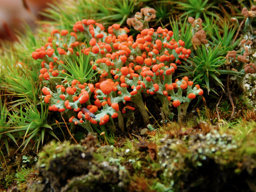
Cladonia cristatellaScarlet tips on 1–3 cm stalksGrey-green squamulesLogs, stumps, soil
Herpothallon rubrocinctumNone; the red is a cottony rim and granulesGrey-green crust with white and red zonesBark, southeastern US

Black patches on rock

WhatLookWhere
Hydropunctaria mauraFlat matt black crust, finely cracked like dried mud, with minute black dots (perithecia); hard to removeContinuous band on shore rock at and above high water
LichinaTiny black tufts or cushions, not a flat crustJust above or below the black crust band
Collema and other gelatinous lichensBrittle black lobes when dry, swelling to olive jelly when wetDamp rock, mortar, bark
Oil or tarNo cracked texture or fruiting dots under a lensIrregular, often at the strandline

Photographing a Lichen for Identification

Whether you ask an expert, post to iNaturalist or use an image-recognition app, the photos decide how far an identification can go. Take several:

  1. The whole lichen in place, showing what it grows on (bark, rock, soil, wood) and something for scale.
  2. A sharp close-up of the surface, through a hand lens or in macro mode: soredia vs isidia, white dots vs a white net, cilia, discs.
  3. The underside and edge of a leafy lichen (lift one lobe gently), or the inside of a strand for beards (is there a central cord?).
  4. Wet and dry if you can; many lichens change colour dramatically when wet.
  5. Notes: region, tree species or rock type, sun or shade, and any spot-test results.

What a photo cannot show. Many lichens are separated only by chemistry or by microscopic features (spores, ascus type, internal anatomy). From a good photo, most common lichens can be named to genus and many conspicuous ones to species; pairs such as Parmelia saxatilis and its relatives, or the Cladonia chlorophaea group, need spot tests or lab work. Treat any photo-only species name as provisional.

Ref

Glossary of Terms

Comprehensive reference for all lichen terminology, with photographs of key structures and growth forms.

← Back to Home